
Influence of hydrogel and porous scaffold on the magnetic thermal property and anticancer effect of Fe3O4 nanoparticles


Abstract
Magnetic hyperthermia uses magnetic nanoparticles (MNPs) for conversion of magnetic energy into thermal energy under an alternating magnetic field (AMF) to increase local temperature for ablation of cancer cells. The magnetic thermal capacity of MNPs not only depends on the intrinsic properties of MNPs but is also affected by the microenvironmental matrices surrounding the MNPs. In this study, the influence of agarose hydrogels and gelatin porous scaffolds on the magnetic thermal property and anticancer effect of Fe3O4 nanoparticles (NPs) were investigated with a comparison to free Fe3O4 NPs. Flower-like Fe3O4 NPs were synthesized and embedded in agarose hydrogels and gelatin porous scaffolds. Under AMF irradiation, the free Fe3O4 NPs had the best magnetic thermal properties and the most efficiently increased the local temperature to ablate breast cancer cells. However, the Fe3O4 NPs embedded in agarose hydrogels and gelatin porous scaffolds showed reduced magnetic-thermal conversion capacity, and the local temperature change was decreased in comparison to free Fe3O4 NPs during AMF irradiation. The gelatin porous scaffolds showed a higher inhibitory influence than the agarose hydrogels. The inhibitory effect of agarose hydrogels and gelatin porous scaffolds on magnetic-thermal conversion capacity resulted in a decreased anticancer ablation capacity to breast cancer cells during AMF irradiation. The Fe3O4 NP-embedded gelatin scaffolds showed the lowest anticancer effect. The results suggested that the matrices used to deliver MNPs could affect their performance, and appropriate matrices should be designed to maximize their therapeutic effect for biomedical applications.
Keywords
INTRODUCTION
Magnetic nanoparticles (MNPs) can convert magnetic energy to thermal energy when subjected to alternating magnetic field (AMF)[1,2]. The heat generated by MNPs under AMF irradiation increases the local temperature to ablate cancer cells, known as magnetic hyperthermia (MH)[3-5]. MH has been developed as an effective approach for cancer treatment due to its good biocompatibility and deep strong tissue penetration. This approach has also been combined with radiotherapy, chemotherapy, and immunotherapy to further improve the anticancer effect[6-12]. It is pivotal to increase magnetic-thermal conversion efficiency to achieve a maximized therapeutic effect with the minimized dosage of MNPs.
To achieve high magnetic-thermal conversion efficiency, many studies have reported the optimization of synthesis methods of MNPs[13,14] by controlling their structure and magnetic characteristics, including shape, size, size distribution, dispersion and aggregation state, crystallinity, composition, and magnetic parameters[15-26]. Except for the intrinsic properties of MNPs, their surrounding microenvironmental matrices can affect the magnetic-thermal conversion[27,28]. Incorporation of MNPs in hydrogels has been reported to change their magnetic-thermal conversion property [28-31]. Engelmann et al. immobilized them in acrylamide hydrogels and found that the heating efficiency of MNPs decreased when the hydrogel stiffness increased[28]. Suto et al. compared the influence of polyvinyl alcohol hydrogel and water[31]. The heating efficiency of MNPs dispersed in water was better than that dispersed in polyvinyl alcohol hydrogel, and their specific absorption rate value in hydrogel showed 67% less than that in water[31]. These studies suggest the inhibitory effects of hydrogels on the magnetic-thermal conversion property of MNPs.
In recent years, localized delivery of photothermal nanoparticles (NPs) has been demonstrated as an efficient strategy to accumulate and constrain them in tumors to maximize the photothermal ablation effect while decreasing their side effect[32-37]. Both hydrogels and porous scaffolds are good carriers for the local delivery of photothermal NPs. However, the influence of porous scaffolds on the magnetic-thermal conversion of MNPs remains elusive. It is desirable to compare the influence of aqueous solution, hydrogels[38-45], and porous scaffolds[46-48] on the magnetic-thermal conversion of MNPs to maximize their MH effect.
Based on the above considerations, in this study, different microenvironments of phosphate buffer saline (PBS), hydrogels, and porous scaffolds were used to investigate the magnetic-thermal conversion property and anticancer effect of MNPs under AMF irradiation [Figure 1]. The same concentration of Fe3O4 NPs was used in PBS, agarose hydrogels, and gelatin porous scaffolds to disclose the influence of the different matrices on magnetic thermal effects. Moreover, Fe3O4 NPs at different concentrations were incorporated in the same matrix to study the MNP concentration dependence. The free Fe3O4 NPs in PBS exhibited the best magnetic thermal property, while embedding in agarose hydrogels or gelatin porous scaffolds decreased the temperature change during AMF irradiation. The anticancer effect was investigated in vitro by incubating breast cancer cells (MDA-MB-231-Luc cells) with free Fe3O4 NPs, agarose/Fe3O4 hydrogels, and
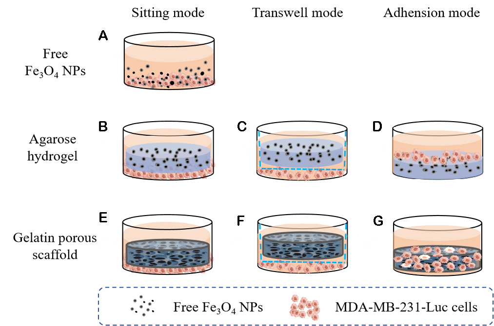
Figure 1. Anticancer experimental scheme of free Fe3O4 NPs (A), agarose/Fe3O4 hydrogels (B-D), and gelatin/Fe3O4 porous scaffolds (E-G). Three modes (sitting, transwell, and adhesion modes) were used to simulate the cells near or far away from or directly adhered to the matrices.
MATERIALS AND METHODS
Materials
Iron (II) chloride tetrahydrate (FeCl2·4H2O, ≥ 99%), iron (III) chloride hexahydrate (FeCl3·6H2O, ≥ 97%), and sodium citrate tribasic dihydrate (≥ 99%) were purchased from Sigma-Aldrich (St. Louis, MO, USA). Diethylene glycol (DEG, 99%), sodium hydroxide (NaOH, 99.99%), acetic acid, ethanol, ethyl acetate,
Synthesis and characterization of Fe3O4 NPs
Fe3O4 NPs were synthesized using Fe (II) and Fe (III) in a mixture solution of DEG and NMDEA
Preparation and characterization of agarose/Fe3O4 hydrogels and gelatin/Fe3O4 scaffolds
The agarose/Fe3O4 hydrogels were prepared by dispersing the citrate-modified Fe3O4 NPs in agarose hydrogels. At first, 0.1 g agarose was dissolved in 5 mL PBS at 110 °C. After the temperature of the agarose solution was cooled to around 40 °C, the citrate-modified Fe3O4 NPs suspension solution was added to prepare 1% agarose solution with Fe3O4 NPs at 5 mg mL-1, 10 mg mL-1, and 20 mg mL-1. Agarose hydrogels without Fe3O4 NPs were prepared as a control. After vortexing, the mixture was added into cylinder molds
The porous scaffolds of gelatin and Fe3O4 NPs were prepared by using ice particulate porogen methods[51-61]. Firstly, ice particulates with a diameter of 250-355 μm were obtained by spraying Milli-Q water into liquid nitrogen and sieved in a low-temperature chamber. Gelatin solution (8%, wt/v) in 70% acetic acid was mixed with the citrate-modified Fe3O4 NP suspension solution (1:1, v/v) to obtain gelatin/Fe3O4 NP mixture solution. Then, the temperature-balanced ice particulates (-4 °C) were added to the gelatin/Fe3O4 NP mixture solution (7:3, wt/v) in a -4 °C chamber, and the final concentrations of Fe3O4 NPs in the mixture solution were 5, 10, and 20 mg cm−3, respectively. The mixture was transformed into a silicone frame and frozen at -20 °C for 12 h and -80 °C for 4 h. Then, the lyophilized constructs were cross-linked by
Magnetic thermal property of Fe3O4 NPs in different matrices
The magnetic thermal properties of free Fe3O4 NPs, agarose/Fe3O4 hydrogels, and gelatin/Fe3O4 porous scaffolds were investigated under AMF irradiation. First, 300 μL of free Fe3O4-5, Fe3O4-10, and Fe3O4-20 solutions were added in 0.5 mL Eppendorf tubes, respectively. The samples were placed in the center of Double H CoilSets AMF (Frequency: 373.6 kHz; Field intensity: 130 Gauss) for 10 min using a D5 series machine (nB nanoScale BioMagnetics, Zaragoza, Spain). Subsequently, the IR1 thermal imaging system (nB nanoScale Biomagnetics, Zaragoza, Spain) was used to record the temperature change of different samples. Triplicate samples were used for each measurement.
Similarly, 300 μL of aqueous agarose solution without or with 5 mg mL-1, 10 mg mL-1, and 20 mg mL-1 of
The gelatin/Fe3O4 porous scaffolds were molded into cylinder discs (Φ10 mm × H4 mm) and hydrated with pure water (300 μL/disc) in silicone frames. The gelatin/Fe3O4 porous scaffold discs were exposed to AMF for 10 min (Frequency: 373.6 kHz; Field intensity: 130 Gauss), and the temperature change was measured. Triplicate samples were used for each measurement.
Anticancer effect of Fe3O4 NPs in different matrices
The anticancer effect of free Fe3O4 NPs, agarose/Fe3O4 hydrogels, and gelatin/Fe3O4 porous scaffolds was investigated by incubating breast cancer cells in the different matrices under AMF irradiation[55-61]. Three culture modes were used to simulate the cells directly adhered to, near, or far away from the matrices [Figure 1]. The cells were seeded in wells of cell culture plates, and then free Fe3O4 NPs, agarose/Fe3O4 hydrogels, or gelatin/Fe3O4 porous scaffolds were added to the adhered cells. The hydrogels and porous scaffolds were sitting on the adhered cells (sitting mode), which simulated the cells near the matrices. The second mode was a transwell mode by seeding cells in the bottom wells of the transwell plates and placing the hydrogels and porous scaffolds in the inserts, which simulated the cells far away from the matrices. The third mode was an adhesion mode by seeding cells on the hydrogels or in the porous scaffolds to allow the cells to adhere to the hydrogels or in the pores of the porous scaffolds, which simulated the cells directly adhering to the matrices.
Anticancer effect of free Fe3O4 NPs
The free Fe3O4 NPs could only be added in the culture medium. Therefore, only the sitting mode was used for investigating the anticancer effect of free Fe3O4 NPs. The sub-cultured MDA-MB-231-Luc cells were harvested and resuspended in a culture medium at a concentration of 2.5 × 10⁵ cells mL−1. A 200 μL cell suspension solution was seeded in the wells of a 48-well plate (5 × 10⁴ cells well-1). After culture in a humidified incubator (5% CO2, 37 °C) for 24 h, the culture medium was removed, and another 200 μL fresh culture medium was added. Then, 300 μL medium, without or with free Fe3O4-5, free Fe3O4-10 and free
Anticancer effect of agarose/Fe3O4 hydrogels
The three culture modes were used for the investigation of the anticancer effect of the agarose/Fe3O4 hydrogel. For the sitting mode, the MDA-MB-231-Luc cells were seeded and cultured in the wells of a 48-well plate, as mentioned above. After the culture medium was changed with another 200 μL fresh medium, the agarose and agarose/Fe3O4 hydrogel discs (Φ10 mm × H4 mm) were placed on the cells. After co-incubation for 2 h, the wells were exposed to AMF (frequency: 373.6 kHz of; field intensity: 130 Gauss) for 10 min. Cell viability was investigated by live/dead staining and WST-1 assay before and after AMF irradiation. Triplicate samples were used for each measurement.
For the transwell mode, the MDA-MB-231-Luc cells were seeded in the centers of the wells of 24-well plate by using donut-shaped silicone rings (inner diameter 10 mm, outer diameter: 16 mm, height: 2 mm). The seeded cell number was the same (5 × 10⁴ cells well-1). The agarose/Fe3O4 hydrogel discs were placed on the transwell inserts and co-cultured with the cells on the bottom wells. The transwell plates containing cells and discs were irradiated by AMF (frequency: 373.6 kHz; magnetic field: 130 Gauss) for 10 min. Before and after AMF irradiation, cell viability was investigated, as mentioned above. Triplicate samples were used for each measurement.
For the adhesion mode, the agarose/Fe3O4 hydrogel discs (Φ10 mm × H4 mm) were put in the wells of a 48-well plate. Subsequently, 200 μL of cell suspension solution was seeded on the agarose/Fe3O4 hydrogel discs. After 2 h incubation, the plates were exposed to AMF (frequency: 373.6 kHz; magnetic field: 130 Gauss) for 10 min, and cell viability was investigated, as mentioned above. Triplicate samples were used for each measurement.
Anticancer effect of gelatin/Fe3O4 porous scaffolds
The three culture modes were also used for the investigation of the anticancer effect of the gelatin and gelatin/Fe3O4 porous scaffolds. All the experiment procedures were the same as those of agarose/Fe3O4 hydrogel discs by using the porous scaffold discs (Φ10 mm × H4 mm). Triplicate samples were used for each measurement.
RESULTS
Characterization of Fe3O4 NPs
The morphology and size of the citrate-modified Fe3O4 NPs were characterized by TEM. As shown in Figure 2A-C, the NPs displayed a flower-like shape, which should have a good magnetic-thermal conversion capacity for MH. They had an average size of 30.8 ± 5.7 nm from the TEM images. The hydrodynamic size of the citrate-modified Fe3O4 NPs was measured in aqueous solution by DLS, and the hydrodynamic size was 108.5 ± 28.5 nm [Figure 2D].

Figure 2. TEM images of citrate-modified Fe3O4 NPs at low (A), middle (B), and high magnifications (C). Hydrodynamic size distribution of citrate-modified Fe3O4 NPs (D).
Characterization of agarose/Fe3O4 hydrogels and gelatin/Fe3O4 porous scaffolds
The agarose/Fe3O4 hydrogel discs are shown in Figure 3A. As the concentration of Fe3O4 NPs increased, the appearance of agarose/Fe3O4 hydrogel changed from transparent to black. SEM observation of the lyophilized agarose/Fe3O4 hydrogel discs showed that the agarose hydrogels with different amounts of Fe3O4 NPs had similar structures [Figure 3B]. They had spindle-shaped pores. The gelatin porous scaffold without Fe3O4 NPs was white, while the gelatin/Fe3O4 porous scaffolds became gray (gelatin/Fe3O4-5), dark gray (gelatin/Fe3O4-10), and black (gelatin/Fe3O4-20) [Figure 3C]. The gelatin and gelatin/Fe3O4 porous scaffolds had the same pore structures. They had large spherical pores that were surrounded by some small pores [Figure 3D]. The large spherical pores were controlled by the ice particulates that were used as a porogen material. The results indicated that the embedding of Fe3O4 NPs did not affect the pore structures of hydrogels and porous scaffolds.
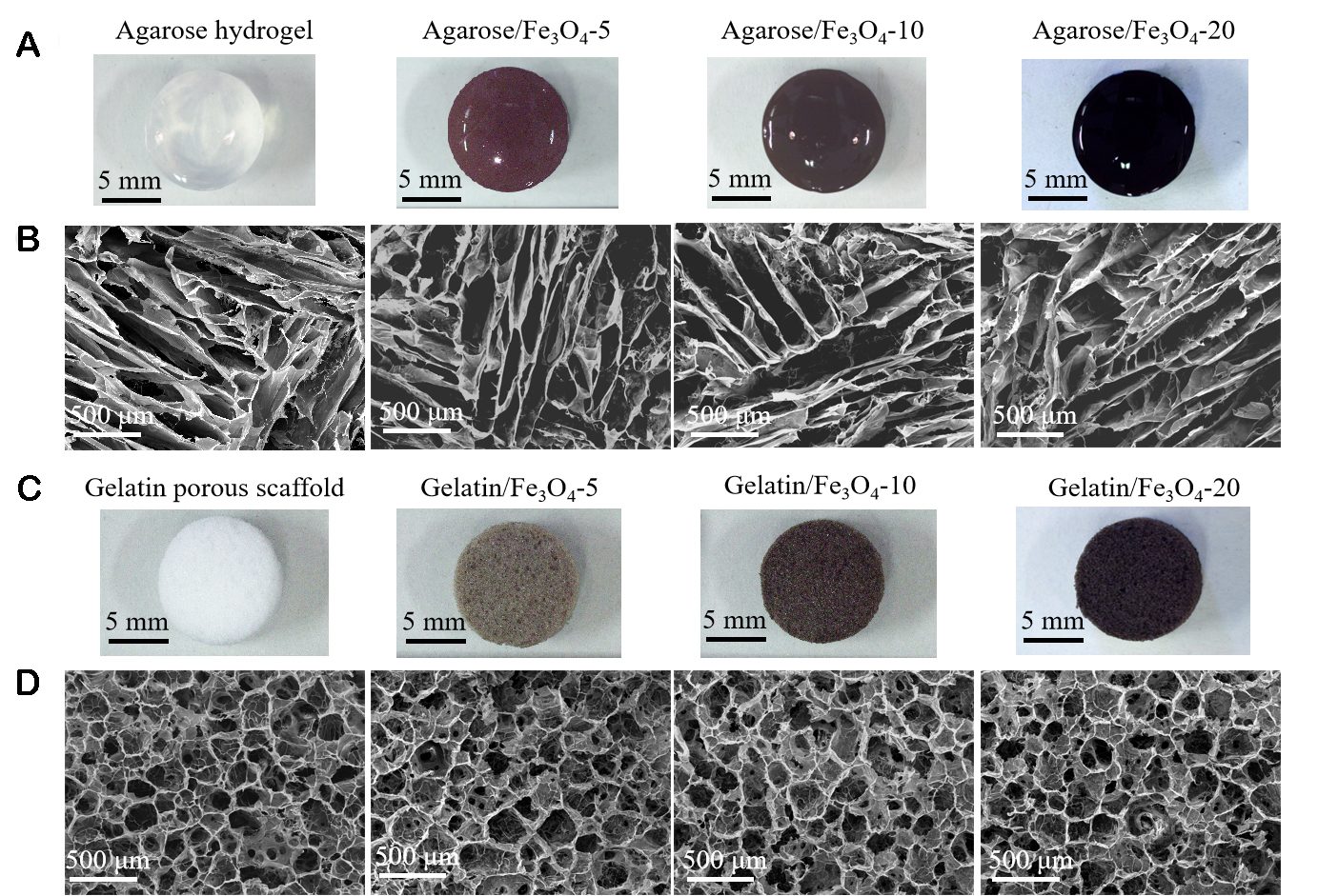
Figure 3. Gross appearances (A) and SEM images (B) of agarose and agarose/Fe3O4 hydrogels with different concentrations of citrate-modified Fe3O4 NPs. Gross appearances (C) and SEM images (D) of gelatin and gelatin/Fe3O4 porous scaffold with different concentrations of citrate-modified Fe3O4 NPs.
Magnetic thermal property of Fe3O4 NPs in different matrices
The magnetic thermal properties of Fe3O4 NPs in PBS, agarose hydrogels, and gelatin porous scaffolds were investigated by applying AMF (frequency: 373.6 kHz; field: 130 Gauss) for 10 min, and the results are shown in Figure 4 and Table 1. The temperature of PBS, agarose hydrogels, and gelatin porous scaffolds without Fe3O4 NPs had no obvious change after AMF irradiation [Figure 4A and Table 1]. The results suggested that PBS, agarose hydrogels, and gelatin porous scaffolds had no magnetic-thermal conversion capacity in the absence of Fe3O4 NPs. When Fe3O4 NPs were added to PBS, hydrogels, and porous scaffolds, the temperature change significantly increased under AMF irradiation.

Figure 4. Heating curves of PBS, agarose hydrogels, and gelatin porous scaffolds without Fe3O4 NPs (A) and containing 5 mg mL-1 Fe3O4 NPs (B), 10 mg mL-1 Fe3O4 NPs (C), and 20 mg mL-1 Fe3O4 NPs (D) during AMF irradiation.
Magnetic thermal property of Fe3O4 NPs in different matrices under AMF irradiation [Mean ± SD (n = 3)]
| Sample | ΔT of PBS (°C) | ΔT of agarose hydrogel (°C) | Percentage compared to free NPs | ΔT of gelatin porous scaffold (°C) | Percentage compared to free NPs |
| No Fe3O4 NPs | 0.7 ± 0.2 | 1.0 ± 0.2 | / | 0.8 ± 0.2 | / |
| Fe3O4-5 mg mL-1 | 24.1 ± 1.7 | 14.0 ± 0.3 | 58.1% | 5.2 ± 0.3 | 21.6% |
| Fe3O4-10 mg mL-1 | 38.3 ± 1.1 | 22.8 ± 1.7 | 59.5% | 9.1 ± 0.5 | 23.8% |
| Fe3O4-20 mg mL-1 | 65.7 ± 1.4 | 33.8 ± 1.0 | 51.5% | 13.2 ± 0.4 | 20.1% |
The temperature change increased with the irradiation time and became plateau after 10 min AMF irradiation [Figure 4B-D]. The temperature change of free Fe3O4-5, agarose/Fe3O4-5, and
The free Fe3O4 NPs in PBS showed the highest temperature change. The temperature change was reduced to 51.5%-59.5% when the Fe3O4 NPs were embedded in agarose hydrogels. The temperature change was further decreased to 20.1%-23.8% when the Fe3O4 NPs were embedded in gelatin porous scaffolds. The results indicated that the matrix where Fe3O4 NPs were embedded could significantly affect the magnetic-thermal conversion property of Fe3O4 NPs.
Anticancer effect of free Fe3O4 NPs
MH uses the Fe3O4 NPs to absorb and convert magnetic energy to heat and raise the local temperature, thereby killing the cancer cells. In this study, MDA-MB-231-Luc cells were cultured in a culture medium supplemented with free Fe3O4 NPs under different concentrations. Cell viability before and after AMF irradiation was investigated by live/dead staining and WST-1 assay [Figure 5]. Before AMF irradiation, almost all the cells were alive [Figure 5A]. After AMF irradiation, the cells cultured without Fe3O4 NPs were still alive, while almost all the cells cultured with Fe3O4-5, Fe3O4-10, and Fe3O4-20 were dead. The results indicated that the cells cultured with free Fe3O4 NPs at a concentration of 5, 10, and 20 mg mL-1 were completely killed after AMF irradiation. Some of the dead cells cultured with 20 mg mL-1L free Fe3O4 NPs detached from the culture wells.
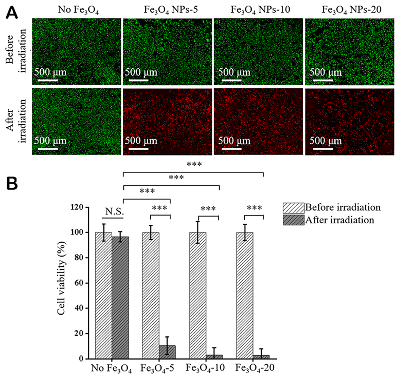
Figure 5. Anticancer effect of free Fe3O4 NPs. Live/dead staining of MDA-MB-231-Luc cells cultured without or with free Fe3O4 NPs before and after AMF irradiation (live cell: green fluorescence, dead cells: red fluorescence) (A). Quantified cell viability during culture without or with free Fe3O4 NPs before and after AMF irradiation (B). Cell viability was normalized to that cultured with PBS without free Fe3O4 NPs before AMF irradiation. Data are the mean ± SD (n = 3). Significant difference: ***P < 0.001. N.S. : no significant difference.
The WST-1 assay showed that the cells cultured without or with free Fe3O4 NPs had the same high viability before AMF irradiation [Figure 5B]. After AMF irradiation, the viability of cells cultured without free Fe3O4 NPs did not change significantly, while the viability of cells cultured with free Fe3O4 NPs significantly decreased after AMF irradiation. Viability of cells cultured with 5, 10, and 20 mg mL-1 free Fe3O4 NPs decreased to 10.3% ± 6.9%, 3.1% ± 5.8%, and 2.7% ± 5.2%, respectively. All the live/dead staining and WST-1 assay results indicated that the breast cancer cells could be killed by the free Fe3O4 NPs under AMF irradiation. A higher concentration of free Fe3O4 NPs resulted in a higher killing effect. The killing effect of free Fe3O4 NPs should be due to the high temperature generated by free Fe3O4 NPs during AMF irradiation [Figure 4]. A higher concentration of the Fe3O4 NPs could generate higher temperatures and enhance the killing efficiency.
Anticancer effect of agarose/Fe3O4 hydrogels
The interaction between cells and agarose/Fe3O4 hydrogels should be different from that between cells and free Fe3O4 NPs. The cells could be near the hydrogels without adhesion to the hydrogels. The cells could also be far away from the hydrogels or directly adhere to the hydrogels. To simulate these interactions between the breast cancer cells and hydrogels, three culture modes were used to investigate the MH anticancer effect of the agarose/Fe3O4 hydrogels. When the MDA-MB-231-Luc cells were cultured near the agarose/Fe3O4 hydrogel discs (sitting mode), most of the cells near the agarose/Fe3O4-5 hydrogel discs were dead, and cell viability decreased significantly after AMF irradiation [Figure 6A and B]. Almost all the cells near the agarose/Fe3O4-10 and agarose/Fe3O4-20 were dead, and their viability further decreased to the lowest level. The cell viability near agarose/Fe3O4-5, agarose/Fe3O4-10, and agarose/Fe3O4-20 decreased to 11.3% ± 4.8%,

Figure 6. Anticancer effect of agarose/Fe3O4 hydrogels. Live/dead staining of MDA-MB-231-Luc cells cultured with the agarose/Fe3O4 hydrogel discs via sitting (A), transwell (C), and adhesion (E) modes before and after AMF irradiation (live cell: green fluorescence, dead cells: red fluorescence). Quantified cell viability during culture without or with agarose/Fe3O4 hydrogel discs via sitting (B), transwell (D), and adhesion (F) modes before and after AMF irradiation. Cell viability was normalized to that cultured with agarose hydrogel without free Fe3O4 NPs before AMF irradiation. Data are the mean ± SD (n = 3). Significant difference: *P < 0.05,
When the breast cancer cells were cultured far away from the agarose/Fe3O4 hydrogel discs (transwell mode), the agarose/Fe3O4 hydrogels with a high concentration of Fe3O4 NPs (agarose/Fe3O4-10 and agarose/Fe3O4-20) could kill almost all the cells and significantly decrease cell viability to very low level by AMF irradiation [Figure 6C and D]. However, only a small number of cells cultured with the
The breast cancer cells adhered on the agarose/Fe3O4 hydrogel discs (adhesion mode) were most efficiently killed by AMF irradiation. Almost all the cells cultured with all the agarose/Fe3O4 hydrogel discs were dead, and their viability was the lowest compared to the sitting and transwell modes [Figure 6E and F]. Viability of breast cancer cells adhered on the agarose/Fe3O4-5, agarose/Fe3O4-10, and agarose/Fe3O4-20 decreased to 7.1% ± 1.2%, 2.7% ± 4.6%, and 2.3% ± 6.5%, respectively, after AMF irradiation [Figure 6F]. This should be due to the direct heating effect of the cells by the agarose/Fe3O4 hydrogels when the cells adhered to the hydrogels.
Anticancer effect of gelatin/Fe3O4 porous scaffolds
The anticancer effect of gelatin/ Fe3O4 porous scaffolds was investigated by using the same methods as those used for the agarose/Fe3O4 hydrogels [Figure 7]. The cells cultured with the gelatin porous scaffold before and after AMF irradiation and with the gelatin/Fe3O4 porous scaffolds before AMF irradiation were alive with high viability. However, after AMF irradiation, some dead cells were observed in the gelatin/Fe3O4-5 and gelatin/Fe3O4-10, and almost all the cells were dead in the gelatin/Fe3O4-20 when the cells were cultured near the scaffolds [Figure 7A] or adhered in the scaffolds [Figure 7E]. When the cells were cultured far away from the scaffolds, a small part of the cells cultured with the gelatin/Fe3O4-20 were killed [Figure 7C]. After AMF irradiation, the viability of breast cancer cells cultured with gelatin/Fe3O4-5, gelatin/Fe3O4-10, and gelatin/Fe3O4-20 was 94.2% ± 9.1%, 80.3% ± 7.8%, and 6.2% ± 5.2% in sitting modes [Figure 7B],
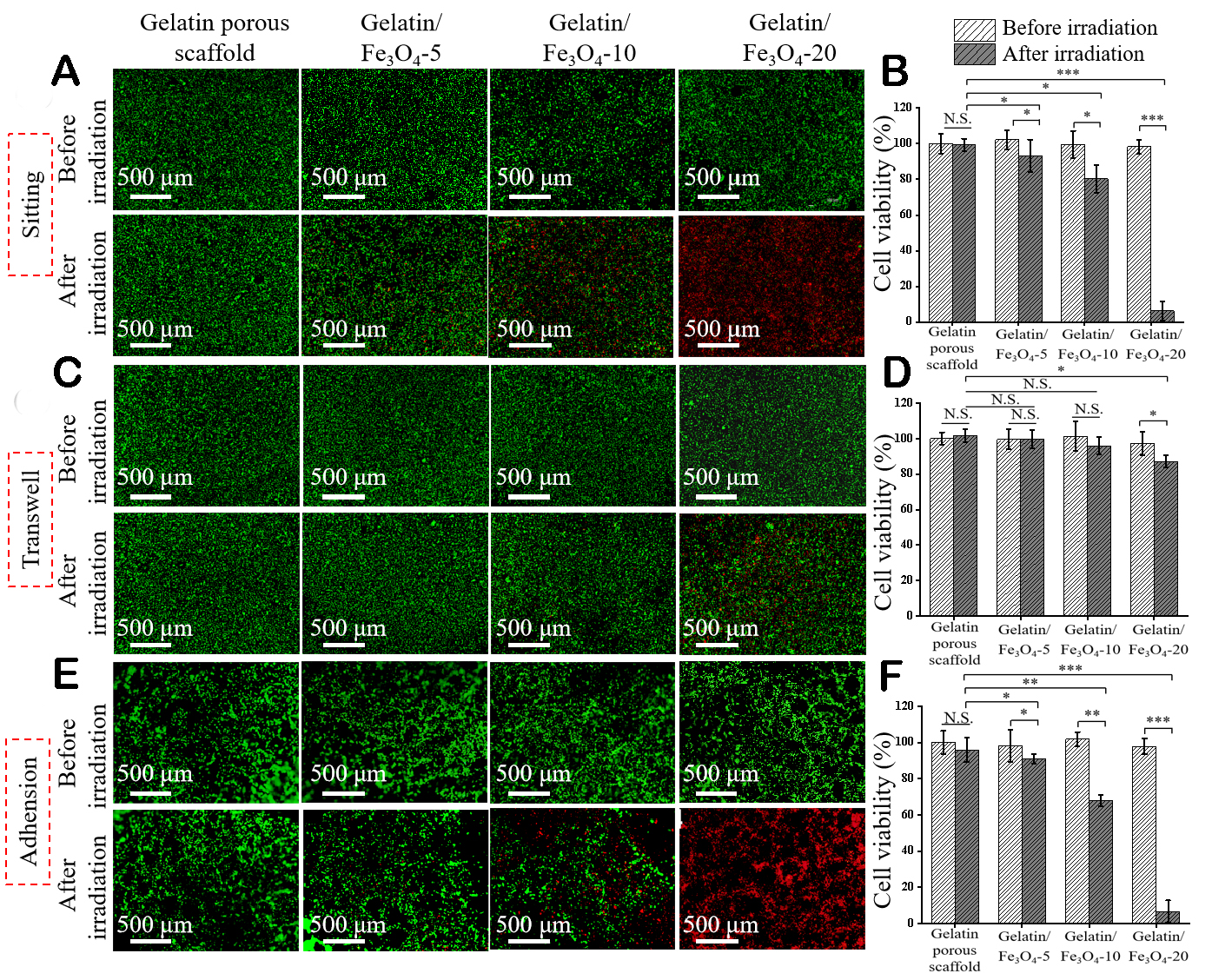
Figure 7. Anticancer effect of gelatin/Fe3O4 porous scaffolds. Live/dead staining of MDA-MB-231-Luc cells cultured with gelatin/Fe3O4 scaffold discs via sitting (A), transwell (C), and adhesion (E) modes before and after AMF irradiation (live cells: green fluorescence, dead cells: red fluorescence). Quantified cell viability after culture with gelatin/Fe3O4 scaffold discs via sitting (B), transwell (D), and adhesion (F) modes before and after AMF irradiation. Cell viability was normalized to that cultured with gelatin porous scaffold without free Fe3O4 NPs before AMF irradiation. Data are the mean ± SD (n = 3). Significant difference: *P < 0.05, **P < 0.01, ***P < 0.001. N.S. : no significant difference.
The above results indicated that the breast cancer cells could be killed by either free Fe3O4 NPs,
DISCUSSION
Fe3O4 NPs have been widely used for MH because of their excellent magnetic-thermal conversion property. Investigation of the influence of matrices surrounding Fe3O4 NPs on the magnetic-thermal conversion property and anticancer effect of Fe3O4 NPs is important for the biomedical application of Fe3O4 NPs to maximize their therapeutic effect. In this study, the magnetic-thermal conversion property and anticancer effect of free Fe3O4 NPs and Fe3O4 NPs embedded in agarose hydrogels and gelatin porous scaffolds were compared because agarose hydrogels and gelatin porous scaffolds have been frequently used to embed therapeutic drugs and NPs. During AMF irradiation, the temperature change of free Fe3O4 NPs,
The matrices used to embed the Fe3O4 NPs could affect the magnetic thermal properties of Fe3O4 NPs. The free Fe3O4 NPs showed the best magnetic thermal properties. Embedding in hydrogels or porous scaffolds decreased the temperature change of Fe3O4 NPs during AMF irradiation. The temperature change of Fe3O4 NPs in agarose hydrogels and porous scaffolds was decreased to 51.5%-59.5% and 20.1%-23.8% of that of free Fe3O4 NPs, respectively. The results of Fe3O4 NPs embedded in hydrogels were the same as the previously reported influence on their magnetic thermal property. Embedding in porous scaffolds further decreased the magnetic-thermal conversion capacity of Fe3O4 NPs.
The matrix influence should be due to the variation of Brownian relaxation of Fe3O4 NPs in the matrices. The heat generation mechanism of MNPs exposed to AMF includes Néel relaxation and Brownian relaxation[62]. Néel relaxation refers to the heating due to the energy loss produced by the rotation of individual magnetic moments within the MNPs under AMF irradiation, and Brownian relaxation refers to the rotation of entire MNPs to produce heat[63,64]. The Néel relaxation of Fe3O4 NPs in the hydrogels and porous scaffolds might not change. However, the matrices should affect the Brownian relaxation of Fe3O4 NPs. The Brownian relaxation of Fe3O4 NPs under AMF irradiation should be partially suppressed in the agarose hydrogel, leading to a decreased heating effect of Fe3O4 NPs. When the Fe3O4 NPs were embedded in gelatin porous scaffolds, the Fe3O4 NPs were tightly constrained in the gelatin fibers, and the Brownian relaxation of Fe3O4 NPs should be heavily inhibited. Therefore, the Fe3O4 NPs in the gelatin porous scaffolds generated heat predominantly through Néel relaxation.
Due to the influence of matrices on the magnetic thermal property of Fe3O4 NPs, the anticancer effect of
Furthermore, the interaction between the breast cancer cells and the Fe3O4 NPs could affect the killing effect. The free Fe3O4 NPs could be uptaken by breast cancer cells and generate heat inside the cells under AMF irradiation. When the Fe3O4 NP-embedded agarose hydrogels were applied, the breast cancer cells could be adhered to the hydrogel, near the hydrogels, or far away from the hydrogels. For the Fe3O4 NP-embedded gelatin porous scaffolds, breast cancers could enter the scaffolds and adhere in the scaffolds, near the scaffolds, or far away from the scaffolds. Three cultured models (sitting, transwell, and adhesion modes) were used to simulate the interaction between the cells and matrices. The Fe3O4 NPs embedded in the agarose hydrogels and gelatin porous scaffolds should be less or not uptaken by the cells if the hydrogels and scaffolds were not degraded. Therefore, the heat should be generated by the scaffolds and then transmitted to the breast cancer cells for ablation. The breast cancer cells adhered to the hydrogels or in the scaffolds were most efficiently ablated. The cells far away from the hydrogels and porous scaffolds were less affected. The results should be due to the heat transmission effect of the hydrogels and porous scaffolds.
CONCLUSION
In this study, the magnetic thermal property and ablation effect of free Fe3O4 NPs and Fe3O4 NPs embedded in agarose hydrogels and gelatin porous scaffolds were investigated to elucidate the influence of microenvironmental matrices on these properties. The flower-like Fe3O4 NPs were embedded in agarose hydrogels and gelatin porous scaffolds. Their magnetic thermal property and anticancer effects were compared with those of free Fe3O4 NPs. Under AMF irradiation, the free Fe3O4 NPs showed the highest temperature increase. Embedding in agarose hydrogels and gelatin porous scaffolds inhibited the heating capacity of Fe3O4 NPs and decreased the temperature change. The gelatin porous scaffolds had the highest inhibitory influence. The anticancer effect of Fe3O4 NPs was also dependent on the matrices. The free Fe3O4 NPs could most efficiently kill breast cancer cells under AMF irradiation. However, the ablation capacity of Fe3O4 NPs embedded in the agarose hydrogels and gelatin porous scaffolds significantly decreased under AMF irradiation compared to that of free Fe3O4 NPs. The reduced killing capacity of Fe3O4 NPs in agarose hydrogels and gelatin porous scaffolds was due to the inhibitory effect of the matrices on their magnetic thermal property. These results suggested that the matrices surrounding MNPs could affect the magnetic thermal property of MNPs and, therefore, affect their ablation capacity to cancer cells. The results should provide useful information for the design and application of MNPs for biomedical applications.
DECLARATIONS
Authors’ contributions
Made substantial contributions to the conception and design of the study and performed data analysis and interpretation: Wang M, Sun R, Kawazoe N, Chen G
Performed data acquisition and provided administrative, technical, and material support: All authors
Availability of data and materials
Data will be made available upon request.
Financial support and sponsorship
This research was supported by the JSPS KAKENHI Grant Number 19H04475.
Conflicts of interest
All authors declared that there are no conflicts of interest.
Ethical approval and consent to participate
Not applicable.
Consent for publication
Not applicable.
Copyright
© The Author(s) 2023.
REFERENCES
1. Tong S, Quinto CA, Zhang L, Mohindra P, Bao G. Size-dependent heating of magnetic iron oxide nanoparticles. ACS Nano 2017;11:6808-16.
2. Hervault A, Thanh NT. Magnetic nanoparticle-based therapeutic agents for thermo-chemotherapy treatment of cancer. Nanoscale 2014;6:11553-73.
3. Cao Z, Wang D, Li Y, et al. Effect of nanoheat stimulation mediated by magnetic nanocomposite hydrogel on the osteogenic differentiation of mesenchymal stem cells. Sci China Life Sci 2018;61:448-56.
4. Liu X, Zheng J, Sun W, et al. Ferrimagnetic vortex nanoring-mediated mild magnetic hyperthermia imparts potent immunological effect for treating cancer metastasis. ACS Nano 2019;13:8811-25.
5. Liu X, Zhang Y, Wang Y, et al. Comprehensive understanding of magnetic hyperthermia for improving antitumor therapeutic efficacy. Theranostics 2020;10:3793-815.
6. Maier-Hauff K, Ulrich F, Nestler D, et al. Efficacy and safety of intratumoral thermotherapy using magnetic iron-oxide nanoparticles combined with external beam radiotherapy on patients with recurrent glioblastoma multiforme. J Neurooncol 2011;103:317-24.
7. Johannsen M, Gneveckow U, Thiesen B, et al. Thermotherapy of prostate cancer using magnetic nanoparticles: feasibility, imaging, and three-dimensional temperature distribution. Eur Urol 2007;52:1653-61.
8. Suriyanto, Ng EY, Kumar SD. Physical mechanism and modeling of heat generation and transfer in magnetic fluid hyperthermia through Néelian and Brownian relaxation: a review. Biomed Eng Online 2017;16:36.
9. Di Corato R, Espinosa A, Lartigue L, et al. Magnetic hyperthermia efficiency in the cellular environment for different nanoparticle designs. Biomaterials 2014;35:6400-11.
10. Balakrishnan PB, Silvestri N, Fernandez-Cabada T, et al. Exploiting unique alignment of cobalt ferrite nanoparticles, mild hyperthermia, and controlled intrinsic cobalt toxicity for cancer therapy. Adv Mater 2020;32:e2003712.
11. Lu N, Huang P, Fan W, et al. Tri-stimuli-responsive biodegradable theranostics for mild hyperthermia enhanced chemotherapy. Biomaterials 2017;126:39-48.
12. Zhang J, Zhao B, Chen S, et al. Near-infrared light irradiation induced mild hyperthermia enhances glutathione depletion and DNA interstrand cross-link formation for efficient chemotherapy. ACS Nano 2020;14:14831-45.
13. Chen S, Zhang Q, Nakamoto T, Kawazoe N, Chen G. Gelatin Scaffolds with controlled pore structure and mechanical property for cartilage tissue engineering. Tissue Eng Part C Methods 2016;22:189-98.
14. Conde-leboran I, Baldomir D, Martinez-boubeta C, et al. A single picture explains diversity of hyperthermia response of magnetic nanoparticles. J Phys Chem C 2015;119:15698-706.
15. de Sousa ME, Carrea A, Mendoza Zélis P, et al. Stress-induced gene expression sensing intracellular heating triggered by magnetic hyperthermia. J Phys Chem C 2016;120:7339-48.
16. Munoz-Menendez C, Conde-Leboran I, Serantes D, Chantrell R, Chubykalo-Fesenko O, Baldomir D. Distinguishing between heating power and hyperthermic cell-treatment efficacy in magnetic fluid hyperthermia. Soft Matter 2016;12:8815-8.
17. Domenech M, Marrero-Berrios I, Torres-Lugo M, Rinaldi C. Lysosomal membrane permeabilization by targeted magnetic nanoparticles in alternating magnetic fields. ACS Nano 2013;7:5091-101.
18. Villanueva A, de la Presa P, Alonso JM, et al. Hyperthermia hela cell treatment with silica-coated manganese oxide nanoparticles. J Phys Chem C 2010;114:1976-81.
19. Baki A, Remmo A, Löwa N, Wiekhorst F, Bleul R. Albumin-coated single-core iron oxide nanoparticles for enhanced molecular magnetic imaging (MRI/MPI). Int J Mol Sci 2021;22:6235.
20. Obaidat IM, Issa B, Haik Y. Magnetic properties of magnetic nanoparticles for efficient hyperthermia. Nanomaterials 2015;5:63-89.
21. Bauer LM, Situ SF, Griswold MA, Samia AC. High-performance iron oxide nanoparticles for magnetic particle imaging - guided hyperthermia (hMPI). Nanoscale 2016;8:12162-9.
22. Coral DF, Zélis PM, Marciello M, et al. Effect of nanoclustering and dipolar interactions in heat generation for magnetic hyperthermia. Langmuir 2016;32:1201-13.
23. Gavilán H, Simeonidis K, Myrovali E, et al. How size, shape and assembly of magnetic nanoparticles give rise to different hyperthermia scenarios. Nanoscale 2021;13:15631-46.
24. Darwish MSA. Effect of carriers on heating efficiency of oleic acid-stabilized magnetite nanoparticles. J Mol Liq 2017;231:80-5.
25. Bordet A, Landis RF, Lee Y, et al. Water-dispersible and biocompatible iron carbide nanoparticles with high specific absorption rate. ACS Nano 2019;13:2870-8.
26. Gonçalves J, Nunes C, Ferreira L, et al. Coating of magnetite nanoparticles with fucoidan to enhance magnetic hyperthermia efficiency. Nanomaterials 2021;11:2939.
27. Cabrera D, Lak A, Yoshida T, et al. Unraveling viscosity effects on the hysteresis losses of magnetic nanocubes. Nanoscale 2017;9:5094-101.
28. Engelmann UM, Seifert J, Mues B, et al. Heating efficiency of magnetic nanoparticles decreases with gradual immobilization in hydrogels. J Magn Magn Mater 2019;471:486-94.
29. Kaczmarek K, Mrówczyński R, Hornowski T, Bielas R, Józefczak A. The effect of tissue-mimicking phantom compressibility on magnetic hyperthermia. Nanomaterials 2019;9:803.
30. Cabrera D, Coene A, Leliaert J, et al. Dynamical magnetic response of iron oxide nanoparticles inside live cells. ACS Nano 2018;12:2741-52.
31. Suto M, Hirota Y, Mamiya H, et al. Heat dissipation mechanism of magnetite nanoparticles in magnetic fluid hyperthermia. J Magn Magn Mater 2009;321:1493-6.
32. Dong S, Chen Y, Yu L, Lin K, Wang X. Magnetic Hyperthermia-synergistic H2O2 self-sufficient catalytic suppression of osteosarcoma with enhanced bone-regeneration bioactivity by 3D-printing composite scaffolds. Adv Funct Mater 2020;30:1907071.
33. Bigham A, Aghajanian AH, Saudi A, Rafienia M. Hierarchical porous Mg2SiO4-CoFe2O4 nanomagnetic scaffold for bone cancer therapy and regeneration: surface modification and in vitro studies. Mater Sci Eng C Mater Biol Appl 2020;109:110579.
34. Farzin A, Fathi M, Emadi R. Multifunctional magnetic nanostructured hardystonite scaffold for hyperthermia, drug delivery and tissue engineering applications. Mater Sci Eng C Mater Biol Appl 2017;70:21-31.
35. Serio F, Silvestri N, Kumar Avugadda S, et al. Co-loading of doxorubicin and iron oxide nanocubes in polycaprolactone fibers for combining magneto-thermal and chemotherapeutic effects on cancer cells. J Colloid Interface Sci 2022;607:34-44.
36. Eivazzadeh-Keihan R, Pajoum Z, Aghamirza Moghim Aliabadi H, et al. Magnetic chitosan-silk fibroin hydrogel/graphene oxide nanobiocomposite for biological and hyperthermia applications. Carbohydr Polym 2023;300:120246.
37. Sun R, Chen H, Zheng J, et al. Composite scaffolds of gelatin and Fe3O4 nanoparticles for magnetic hyperthermia-based breast cancer treatment and adipose tissue regeneration. Adv Healthc Mater 2023;12:e2202604.
38. Lu C, Zheng J, Yoshitomi T, Kawazoe N, Yang Y, Chen G. How hydrogel stiffness affects adipogenic differentiation of mesenchymal stem cells under controlled morphology. ACS Appl Bio Mater 2023;6:3441-50.
39. Wu H, Liu L, Song L, Ma M, Gu N, Zhang Y. Enhanced tumor synergistic therapy by injectable magnetic hydrogel mediated generation of hyperthermia and highly toxic reactive oxygen species. ACS Nano 2019;13:14013-23.
40. Gao F, Xie W, Miao Y, et al. Magnetic hydrogel with optimally adaptive functions for breast cancer recurrence prevention. Adv Healthc Mater 2019;8:e1900203.
41. Stocke NA, Sethi P, Jyoti A, et al. Toxicity evaluation of magnetic hyperthermia induced by remote actuation of magnetic nanoparticles in 3D micrometastasic tumor tissue analogs for triple negative breast cancer. Biomaterials 2017;120:115-25.
42. Zhang X, Wei P, Wang Z, et al. Herceptin-conjugated DOX-Fe3O4/P(NIPAM-AA-MAPEG) nanogel system for HER2-targeted breast cancer treatment and magnetic resonance imaging. ACS Appl Mater Interfaces 2022;14:15956-69.
43. Lu J, Guo Z, Xie W, et al. Hypoxia-overcoming breast-conserving treatment by magnetothermodynamic implant for a localized free-radical burst combined with hyperthermia. ACS Appl Mater Interfaces 2021;13:35484-93.
44. Jalili NA, Jaiswal MK, Peak CW, Cross LM, Gaharwar AK. Injectable nanoengineered stimuli-responsive hydrogels for on-demand and localized therapeutic delivery. Nanoscale 2017;9:15379-89.
45. Zheng J, Xie Y, Yoshitomi T, Kawazoe N, Yang Y, Chen G. Stepwise proliferation and chondrogenic differentiation of mesenchymal stem cells in collagen sponges under different microenvironments. Int J Mol Sci 2022;23:6406.
46. Sun R, Chen H, Sutrisno L, Kawazoe N, Chen G. Nanomaterials and their composite scaffolds for photothermal therapy and tissue engineering applications. Sci Technol Adv Mater 2021;22:404-28.
47. Zhang W, Chen Y, Li M, et al. A PDA-functionalized 3D lung scaffold bioplatform to construct complicated breast tumor microenvironment for anticancer drug screening and immunotherapy. Adv Sci 2023;10:e2302855.
48. Chen H, Sun R, Zeng T, et al. Stepwise photothermal therapy and chemotherapy by composite scaffolds of gold nanoparticles, BP nanosheets and gelatin immobilized with doxorubicin-loaded thermosensitive liposomes. Biomater Sci 2022;10:7042-54.
49. Lartigue L, Hugounenq P, Alloyeau D, et al. Cooperative organization in iron oxide multi-core nanoparticles potentiates their efficiency as heating mediators and MRI contrast agents. ACS Nano 2012;6:10935-49.
50. Hemery G, Keyes AC Jr, Garaio E, et al. Tuning sizes, morphologies, and magnetic properties of monocore versus multicore iron oxide nanoparticles through the controlled addition of water in the polyol synthesis. Inorg Chem 2017;56:8232-43.
51. Sutrisno L, Chen H, Chen Y, et al. Composite scaffolds of black phosphorus nanosheets and gelatin with controlled pore structures for photothermal cancer therapy and adipose tissue engineering. Biomaterials 2021;275:120923.
52. Li J, Zhang J, Chen Y, Kawazoe N, Chen G. TEMPO-conjugated gold nanoparticles for reactive oxygen species scavenging and regulation of stem cell differentiation. ACS Appl Mater Interfaces 2017;9:35683-92.
53. Zheng J, Sun R, Chen H, et al. Morphological dependence of breast cancer cell responses to doxorubicin on micropatterned surfaces. Polymers 2022;14:2761.
54. Chen Y, Kawazoe N, Chen G. Preparation of dexamethasone-loaded biphasic calcium phosphate nanoparticles/collagen porous composite scaffolds for bone tissue engineering. Acta Biomater 2018;67:341-53.
55. Zheng J, Wang Y, Kawazoe N, Yang Y, Chen G. Influences of viscosity on the osteogenic and adipogenic differentiation of mesenchymal stem cells with controlled morphology. J Mater Chem B 2022;10:3989-4001.
56. Sutrisno L, Chen H, Yoshitomi T, Kawazoe N, Yang Y, Chen G. PLGA-collagen-BPNS bifunctional composite mesh for photothermal therapy of melanoma and skin tissue engineering. J Mater Chem B 2022;10:204-13.
57. Ma H, Zhuang H, Zhai D, et al. Xonotlite nanowire-containing bioactive scaffolds for the therapy of defective adipose tissue in breast cancer. Nano Lett 2023;23:7157-65.
58. Chen H, Sun R, Zheng J, Kawazoe N, Yang Y, Chen G. Doxorubicin-encapsulated thermosensitive liposome-functionalized photothermal composite scaffolds for synergistic photothermal therapy and chemotherapy. J Mater Chem B 2022;10:4771-82.
59. Chen H, Wang X, Sutrisno L, et al. Folic acid-functionalized composite scaffolds of gelatin and gold nanoparticles for photothermal ablation of breast cancer cells. Front Bioeng Biotechnol 2020;8:589905.
60. Sutrisno L, Chen H, Yoshitomi T, Kawazoe N, Yang Y, Chen G. Preparation of composite scaffolds composed of gelatin and Au nanostar-deposited black phosphorus nanosheets for the photothermal ablation of cancer cells and adipogenic differentiation of stem cells. Biomater Adv 2022;138:212938.
61. Zhang J, Li J, Kawazoe N, Chen G. Composite scaffolds of gelatin and gold nanoparticles with tunable size and shape for photothermal cancer therapy. J Mater Chem B 2017;5:245-53.
62. Pinheiro IF, Brollo ME, Bassani GS, et al. Effect of viscosity and colloidal stability on the magnetic hyperthermia of petroleum-based nanofluids. Fuel 2023;331:125810.
63. Fortin JP, Gazeau F, Wilhelm C. Intracellular heating of living cells through Néel relaxation of magnetic nanoparticles. Eur Biophys J 2008;37:223-8.
64. Fortin JP, Wilhelm C, Servais J, Ménager C, Bacri JC, Gazeau F. Size-sorted anionic iron oxide nanomagnets as colloidal mediators for magnetic hyperthermia. J Am Chem Soc 2007;129:2628-35.
65. Wang X, Yang Y, Hu X, Kawazoe N, Yang Y, Chen G. Morphological and mechanical properties of osteosarcoma microenvironment cells explored by atomic force microscopy. Anal Sci 2016;32:1177-82.
66. Calzado-Martín A, Encinar M, Tamayo J, Calleja M, San Paulo A. Effect of actin organization on the stiffness of living breast cancer cells revealed by peak-force modulation atomic force microscopy. ACS Nano 2016;10:3365-74.
67. Qi G, Zhang Y, Xu S, et al. Nucleus and mitochondria targeting theranostic plasmonic surface-enhanced raman spectroscopy nanoprobes as a means for revealing molecular stress response differences in hyperthermia cell death between cancerous and normal cells. Anal Chem 2018;90:13356-64.
Cite This Article
Export citation file: BibTeX | RIS
OAE Style
Wang M, Sun R, Chen H, Liu X, Yoshitomi T, Takeguchi M, Kawazoe N, Yang Y, Chen G. Influence of hydrogel and porous scaffold on the magnetic thermal property and anticancer effect of Fe3O4 nanoparticles. Microstructures 2023;3:2023042. http://dx.doi.org/10.20517/microstructures.2023.46
AMA Style
Wang M, Sun R, Chen H, Liu X, Yoshitomi T, Takeguchi M, Kawazoe N, Yang Y, Chen G. Influence of hydrogel and porous scaffold on the magnetic thermal property and anticancer effect of Fe3O4 nanoparticles. Microstructures. 2023; 3(4): 2023042. http://dx.doi.org/10.20517/microstructures.2023.46
Chicago/Turabian Style
Wang, Man, Rui Sun, Huajian Chen, Xiaohan Liu, Toru Yoshitomi, Masaki Takeguchi, Naoki Kawazoe, Yingnan Yang, Guoping Chen. 2023. "Influence of hydrogel and porous scaffold on the magnetic thermal property and anticancer effect of Fe3O4 nanoparticles" Microstructures. 3, no.4: 2023042. http://dx.doi.org/10.20517/microstructures.2023.46
ACS Style
Wang, M.; Sun R.; Chen H.; Liu X.; Yoshitomi T.; Takeguchi M.; Kawazoe N.; Yang Y.; Chen G. Influence of hydrogel and porous scaffold on the magnetic thermal property and anticancer effect of Fe3O4 nanoparticles. Microstructures. 2023, 3, 2023042. http://dx.doi.org/10.20517/microstructures.2023.46
About This Article
Special Issue
Copyright

Data & Comments
Data


 Cite This Article 9 clicks

 Like This Article 3
likes
Like This Article 3
likes


Comments
Comments must be written in English. Spam, offensive content, impersonation, and private information will not be permitted. If any comment is reported and identified as inappropriate content by OAE staff, the comment will be removed without notice. If you have any queries or need any help, please contact us at support@oaepublish.com.